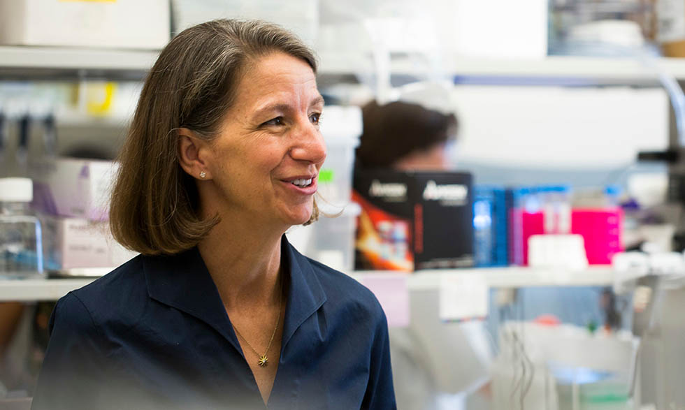
Rebecca Richards-Kortum, Ph.D., of Rice University, has devoted her career to understanding how technology can improve health and save lives. Her recent research focuses on creating affordable screening tools for cervical cancer, the fourth most common cancer among women worldwide.
Imaging technology has helped turn this goal into reality. The technology was developed with support from the National Institute of Biomedical Imaging and Bioengineering and the National Cancer Institute.
Improving cervical cancer detection
There are two main challenges in testing for cervical cancer and human papillomavirus (HPV), the virus that causes cervical cancer: It requires costly tools and extensive lab work.
"Both of these challenges are really important for patients who are medically underserved," Dr. Richards-Kortum notes. "Those could be patients who live in rural areas or poor areas of the U.S. or in low- and middle-income countries around the world."
More than 90% of cervical cancer deaths happen in low- and middle-income countries, according to the World Health Organization. That's where Dr. Richards-Kortum and her colleagues come in.
A portable microscope
They've developed a low-cost fiber optic microscope that allows health care providers to see the same things they would during a tissue biopsy. A biopsy is the most effective way to diagnose cervical cancer.
"We can make this technology for very low cost, it runs on a tablet computer, and it's completely portable and battery powered."
- Rebecca Richards-Kortum, Ph.D.
"We can make this technology for very low cost, it runs on a tablet computer, and it's completely portable and battery powered," she says.
It also requires less training and expertise to use. Usually, women with an abnormal Pap smear have to have a procedure called a colposcopy. During this procedure, a provider takes a small tissue sample from the cervix. The sample is sent to a lab to be examined for cancer cells. With the new technology, providers can take a tissue sample, examine it, and follow up with the patient—all at the same time.
First, a drop of dye is put on the tissue in a woman's cervix. A small fiber-optic probe, about the size of a pencil, is gently placed on the dyed tissue. The probe creates an image of the tissue and cells that it sends back to a "microscope" on the tablet computer, where the provider can review the image for cancer.
Improving follow-up rates
Dr. Richards-Kortum and her colleagues tested the technology in mobile diagnostic vans. They traveled around the Rio Grande Valley in Texas and in Brazil, bringing screening to women where they live.
"It was really exciting to us to see the potential of something not just in a middle-income country like Brazil, but to look right here in our own backyard," she says.
In addition to being affordable and easy to transport, the microscope is also effective. Dr. Richards-Kortum says that during field testing, the technology had a level of accuracy very similar to an expert gynecologist performing a tissue biopsy.
In a clinical trial in Brazil, teams found that easier screenings helped to dramatically improve follow-up rates. "In women who had access to this mobile van, there was almost a 40% increase in the diagnostic follow-up," Dr. Richards-Kortum says.
Dr. Richards-Kortum and her colleagues are also testing the technology with oral cancer.
The future of medical technology
Dr. Richards-Kortum notes that imaging technology has more than just physical health benefits. She has seen how it can improve patients' understanding of their own health.
"It's really interesting to see the power of imaging to help patients better understand the changes that are taking place," she says. "When a provider can point out images and say, 'This is what I see and this is why I'm concerned,' it's very rewarding."
As a leader and mentor in the field of medical technology, Dr. Richards-Kortum is also focused on empowering future generations of bioengineers. She encourages more people to study bioengineering and to take on leadership roles in the field, especially women.
"I think for me, many of my colleagues, and the students we work with, it's an amazing opportunity and privilege to think of how you can use science and engineering to really make people's lives better."


