For centuries, scientists have used small animal models to study everything from how genes work to how diseases develop. And these pint-sized creatures continue to play important roles in scientific discoveries of all kinds, including vision research.
Small animals teach researchers big lessons about how vision works, how it can go wrong (and why), and new ways to treat eye diseases.
How fruit flies transformed human vision research
In the early 1900s, a young graduate student named Mildred Hoge discovered a group of fruit flies with eyes that were unusually small…or missing completely. She mapped the gene responsible for this mutation and named it eyeless. Her later research with fruit flies showed that this gene plays a key role in how their eyes and optic nerves develop.
Almost a century later, scientists discovered that humans have their own version of the eyeless gene, called PAX6. Genetic mutations that affect PAX6 can lead to significant issues such as cataracts and aniridia (a condition that affects the iris of the eye).
NIH-supported researchers continue to study fruit fly development, asking questions like, “What makes an eye an eye?” and “What keeps it from turning into some other body part?”
What makes an eye…an eye?
Justin Kumar, Ph.D., studies and teaches developmental biology at Indiana University, Bloomington. He uses fruit fly models to study how a cell becomes one kind of tissue (like an eye) and not another (like a wing or an antenna). PAX6 provides instructions for making proteins that drive tissue development in the fruit fly eye by turning on "eye" genes and turning off "non-eye" genes. But just changing PAX6 on its own doesn’t let a cell become any type of tissue.
Dr. Kumar discovered that PAX6 proteins work together with another type of protein (called an epigenetic enzyme) to control how DNA is used in cells. This helps cells decide what kind of tissue they will become. He and his team found a way to study this process by turning off or “knocking out” both proteins at the same time. Suddenly, eye tissue can turn into all sorts of things—including a wing!
By looking at the changes to the modified cell, Dr. Kumar can tease out what makes a tissue one type versus another.
“When you knock them out together, it’s this kind of magic bullet, but that’s hidden when you look at them individually,” he explained.
Let’s explore how other NIH-funded vision researchers are using different animal models in their work.
Watch this video to learn more about fruit flies as animal models in vision research.
Studying lens regeneration in newts
Some animals have remarkable abilities that seem almost supernatural. For example, newts can regenerate (regrow) body parts, including tissues of the eye. Katia Del Rio-Tsonis, Ph.D., of Miami University, Ohio, uses a 3D imaging technology called optical coherence tomography (OCT) to watch newts’ lenses regenerate.
“The newt can basically regenerate anything,” she said. “The eye is really a fantastic model, too, because it’s easily manipulated.”
When the lens is removed from a newt’s eye, the immune system clears out any damaged cells and debris. Iris cells transform into new lens cells that grow over time to create a new lens. Eventually, the new lens detaches from the iris and reattaches to the normal connecting tissue. And with OCT, Dr. Del Rio-Tsonis can see how cells and tissues rearrange themselves to rebuild a critical part of the eye.
Discover more about the newt eye lens as a research model in this video.
Learning how zebrafish regrow parts of their eyes could help scientists develop new treatments for blindness
Newts aren’t the only animals with impressive regenerative abilities. Tiny zebrafish can also regrow parts of the eye, including neurons in the eye’s retina (the light-sensitive tissue at the back of the eyeball). Researching this ability could someday help scientists treat blindness in humans.
The eye’s retina is made up of photoreceptor (light-sensitive) cells and nerve cells in the optic nerve (which connects the eye to the brain). In humans, if these cells die, they cannot be replaced. Diseases that damage the retina or optic nerve (such as glaucoma) can lead to permanent blindness.
Zebrafish’s immune systems respond differently than ours. In humans, our immune response causes scar tissue to form, which can stop parts of the retina from responding to light. But when a zebrafish’s retina is damaged, certain cells trigger the affected tissue to regrow and restore vision, a process called “retinal regeneration.”
Daniel Goldman, Ph.D., of the University of Michigan, studies how zebrafish regenerate their eyes. He hopes that this knowledge will lead to treatments that enable humans to regrow neurons in their own eyes. He said that after an optic nerve injury, “[the fish] regain sight…and it happens really fast. Over a two-week period, they’ll reconnect with the brain, and the fish can see again.”
Researching rare diseases with geckos
For clinical scientists like Robert Hufnagel, M.D., Ph.D., animal models can shed light on what’s happening to specific patients who come to the clinic.
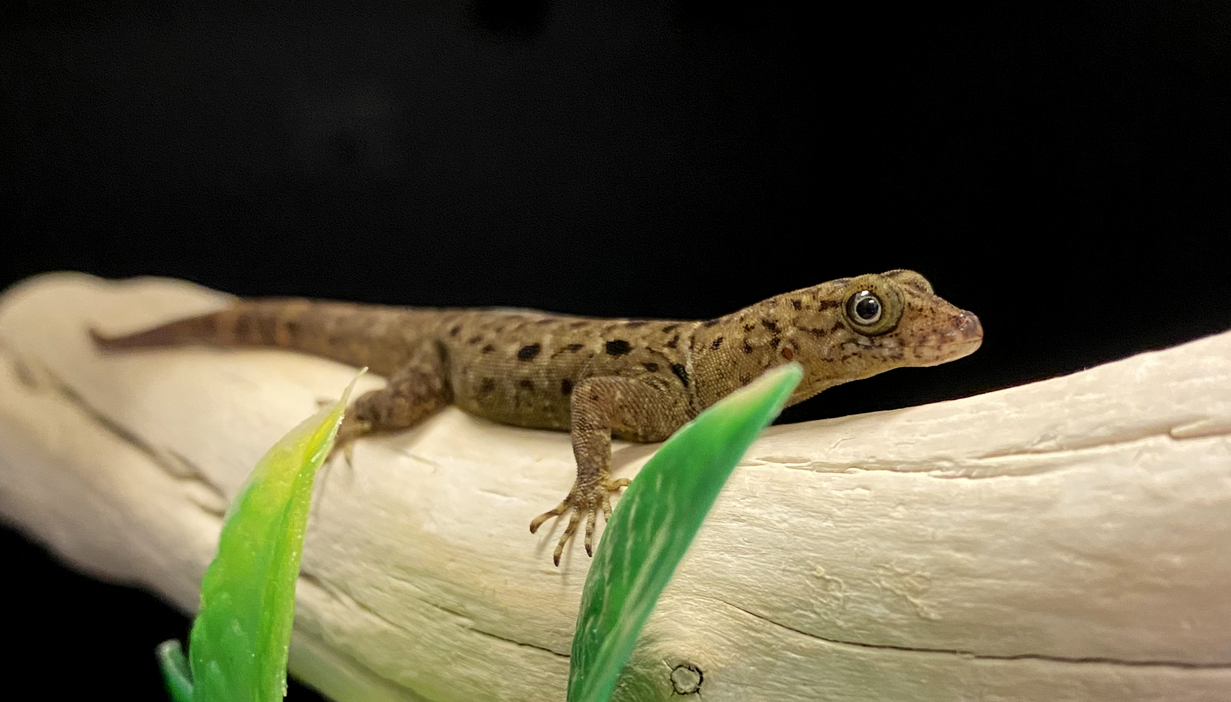
In his lab at the National Eye Institute, Dr. Hufnagel worked with several animal models—including geckos—to study some of the rare diseases he sees in his patients.
“Being able to create relevant animal models to help study these rare diseases is important to our patients,” he explained.
Ashley Rasys, D.V.M, Ph.D., worked as a postdoctoral student in Dr. Hufnagel’s lab. She explained that gecko and human eyes have similar developmental pathways and structures. They also both have a fovea, which is the part of the retina that allows us to see clearly and focus on objects directly in front of us.
Dr. Hufnagel will use a gene-editing tool to introduce changes to the gecko’s DNA that mimic those in his human patients. This will help him learn more about how these genetic changes cause disease. And it will allow him to test new treatments and therapies in the geckos to see how they might work in humans.
*This article was adapted from a longer story by the National Eye Institute. Read the original article to learn more about animal models and the vision researchers that work with them.


