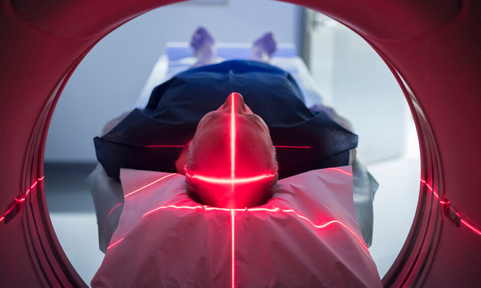
Imaging procedures create pictures of areas inside your body that help your health care provider see whether cancer is present. They can also show if certain cancer treatments are working.
Before getting an imaging procedure, make sure to talk with your health care provider about whether the procedure is necessary and its risks and benefits.
Some imaging procedures can also cause cell damage that leads to cancer. However, the risks of cancer from these medical procedures are very small, and the benefit from having them is almost always greater than the risks.
Imaging tools to help diagnose cancer include:
X-ray:
X-rays use low doses of radiation to create pictures of the inside of your body.
PET (positron emission tomography) imaging:
PET imaging uses a tracer, which is a small amount of radioactive material that flows through your bloodstream. It collects in certain tissues. A scanner then makes 3-D pictures that show where the tracer collects in the body. These scans show how your organs and tissues are working and if disease is present. Your body gets rid of the radioactive substance quickly.
CT (computed tomography) scan:
CT scans use an X-ray machine linked to a computer that takes a series of detailed pictures of your organs. You may first receive a dye or other contrast material (which is injected or ingested) to highlight areas inside the body. Contrast material helps define the appearance of some of the body areas and disease.
Ultrasound:
An ultrasound is a sound wave that people cannot hear. The waves bounce off tissues inside your body. During an ultrasound, a health care provider will use a probe on your skin to detect the echoes and a small scanner to create a picture of them. This picture is called a sonogram.
MRI (magnetic resonance imaging):
MRIs use a strong magnet linked to a computer to make detailed pictures of your body. You may first receive a dye or other contrast material (which is injected or ingested) to highlight areas inside the body. Contrast material helps define the appearance of some of the body areas and disease. Your health care provider can view these pictures on a monitor and print them on film.
Nuclear scan:
Like PET scans, nuclear scans also involve a radioactive tracer that is injected into your bloodstream. This material then collects in certain body tissues. A machine called a scanner detects and makes a computer picture of the body sites where the tracer goes after it is injected. Your body gets rid of the radioactive substance quickly.


